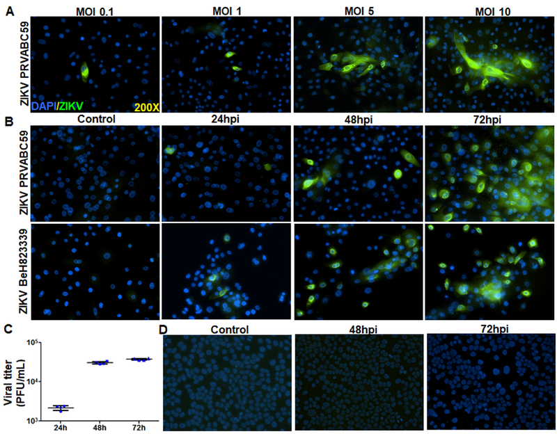
Figure 1. ZIKV infectivity of HCECs: a dose and time course study.
(A) Human Pr. HCEC cells were infected with ZIKV (PRVABC59, a Puerto Rico strain) with various MOI (0.1–10) for 48h. Infected cells were subjected to immunostaining for anti-flavivirus group antigen 4G2, and representative images show the presence of ZIKV (green) and DAPI (blue, a cell nuclear stain). (B) Time course study was performed by infecting Pr. HCEC with two different strains of ZIKV, PRVABC59, and BeH823339 (Brazilian clinical strain) at MOI 5 for the indicated time points, uninfected cells served as control. Immunostaining for anti-flavivirus group antigen 4G2 was performed to detect ZIKV infectivity. (C) Pr. HCECs were infected with ZIKV (strain BeH823339) at MOI 5. The culture supernatant were collected and used for plaque assay on Vero cells. Dot plot represent the plaque forming units (PFU)/ml of conditioned media (n=4) at various time points. (D) A transformed corneal cell line HUCL, were infected with ZIKV, Brazilian strain BeH823339 at MOI 5 and subjected to immunostaining for 4G2 antibody, representative images (n=3) show the presence of ZIKV (green) and DAPI (blue).

