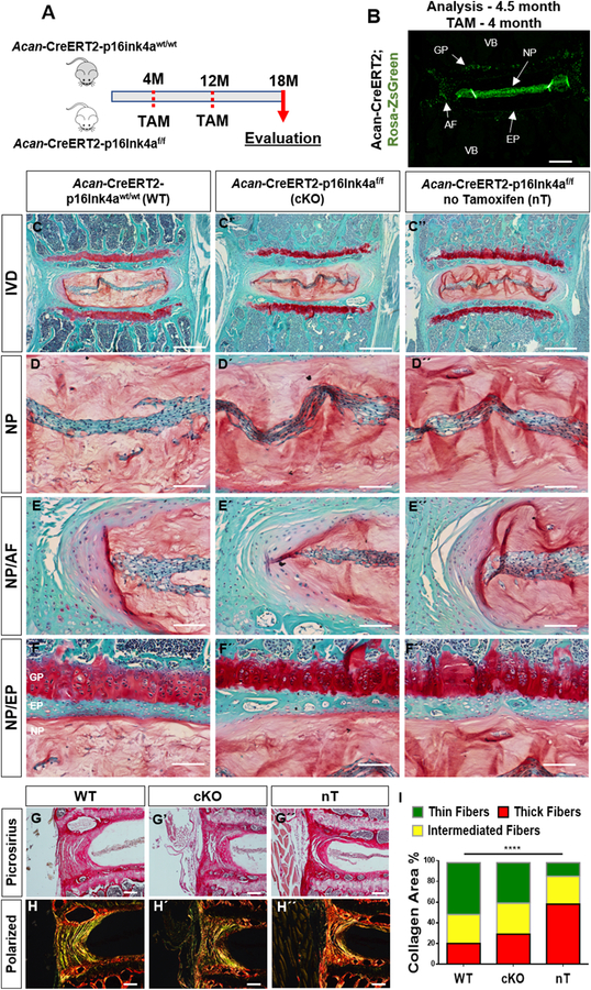
Figure 2. Loss of p16Ink4a in the disc does not protect mice from age-dependent degeneration.
(A) Schematic showing protocol for generation of experimental and control mice. Analysis was performed using tamoxifen treated 18-month-old wild-type (Acan-CreERT2-p16Ink4a wt/wt) and p16Ink4a conditional knock-out (Acan-CreERT2-p16Ink4a f/f) and untreated (Acan-CreERT2-p16Ink4af/f) animals. (B) Specificity of Acan-CreERT2 for targeting different compartments within the intervertebral disc in skeletally mature mice is shown. Acan-Cre showed a high recombination in NP, inner AF, endplate (EP) as well as growth plate (GP), when tamoxifen was administered at 4-month. (C-F’’) There are no noticeable differences in overall intervertebral disc (IVD) architecture or cellular morphology in the tamoxifen treated wild-type (WT) (C-F), conditional knock-out (cKO) (C’-F’), and animals without tamoxifen-treatment (nT) (C’’-F’’) groups as shown by representative histological images. Higher magnification images of NP tissue (D-D”) and tissue interfaces between NP/AF (E-E’’) and NP/EP (F-F’’) show comparable cell morphology and tissue architecture between all the groups. Picrosirius red staining of discs showed similar collagen content among the groups (G-G’’). There were no differences in collagen organization (H-H’’) and distribution of collagen fibers thickness (I) between the WT and cKO mice as seen by quantitative polarized microscopy. Non-treated animals showed higher content of mature collagen than tamoxifen treated WT and cKO animals. P < 0.05, χ2 test, N = 6 mice/group, 4 discs/animal. Scale bar B-C” and G-H´´= 200 µm; Scale bar D-F” = 50 µm.

