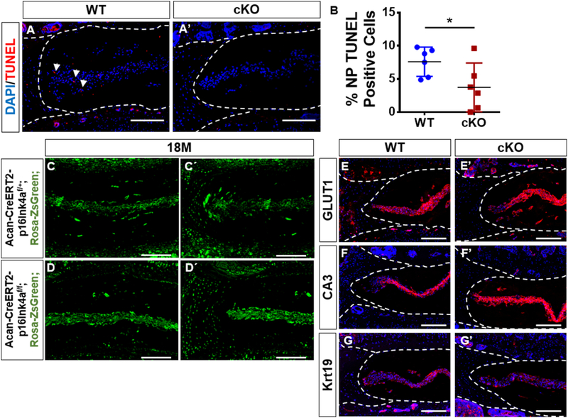
Figure 4. p16Ink4a cKO mice have decreased NP cell death without significant altering cell number and phenotype.
(A-A’’) TUNEL assay showed a decrease in number of dying cells (arrows) in NP compartment of the cKO mouse. (B) There was a decrease in TUNEL positive cells in cKO mice. t-test for normally distributed data, Mann-Whitney test for non-normally distributed data, Shapiro-Wilk normality test was done to check the distribution. NS = not significant; p ≤ 0.05 *; p ≤ 0.01 **; N=6 animals/genotype were analyzed. (C, D’) Fate mapping of disc cells in p16Ink4a cKO and control mice. Tamoxifen treatment of mice (at 4 and 12-months) induced Cre-recombinase activity in aggrecan expressing cells resulting in simultaneous marking of cells by ZsGreen with or without deletion of p16Ink4a. Analysis of these mice at 18-months showed that almost all cells in NP, inner AF and CEP were ZsGreen positive. Cell numbers of ZsGreen positive cells in tissue compartments were comparable between cKO and control animals. (E-G’) Characterization of NP phenotypic markers in p16Ink4a cKO mice. Localization and expression levels of glucose transporter 1 (GLUT1), carbonic anhydrase 3 (CA3) and Keratin19 (Krt19) in the NP were comparable between control and cKO animals. Mann-Whitney test was used for comparing differences between the groups. NS = not significant; p ≤ 0.05 *; p ≤ 0.01 **; N=6 animals/genotype were analyzed. Scale bar = 200µm.

