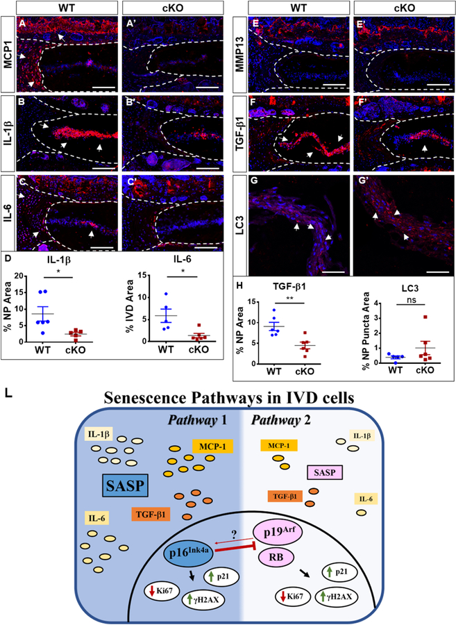
Figure 7. p16Ink4a cKO exhibit decreased expression of major regulators of senescence-associated secretory phenotype (SASP).
(A-C’) cKO animals showed a pronounced decrease in MCP1 (A, A’), IL1β (B, B’) and IL-6 (C, C’) staining compared to control animals (D). (E-E’) There were comparable levels of MMP13 expression between cKO mice and the WT. (F-F’, H) TGF-β1 expression was decreased in cKO discs. Autophagy analysis showed no differences between LC3 Puncta between WT and cKO (G-H). Mann-Whitney test was used for comparing differences between the groups. NS = not significant; p ≤ 0.05 *; N=6 animals/genotype were analyzed. Scale bar = 200 µm. (L) Schematic summarizing contribution of p16Ink4a, p19Arf and RB for senescence status and SASP maintenance in the intervertebral cells.

