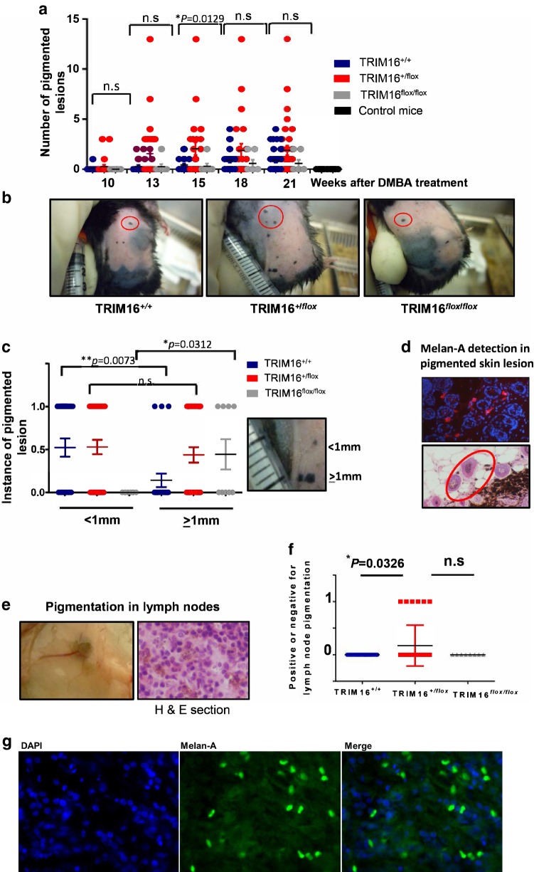
Fig. 3.
TRIM16 knockout mice developed lymph node pigmentation and increased the potential for producing lymph node metastases. a Latency of pigmented cell lesion development was measured by counting the number of pigmented lesions in each cohort of mice every 3 weeks from week 10–21 following a single dose of DMBA tumour initiation treatment. Topical tumour promoting TPA treatment was administered twice weekly. An ANOVA statistical test was performed to assess significance of change in number of lesions present at each time point. b Representative images of wild-type (TRIM16+/+, N = 22), heterozygous (TRIM16+/flox, N = 33) and homozygous (TRIM16flox/flox, N = 8) skin-specific keratinocyte knockout mice with pigmented lesions indicated by red circles. c Melanocytic lesion size was measured as ≥ 1 mm or < 1 mm, 0 indicates no lesion and 1 indicates a lesion. Statistical analysis was performed for each genotype with smaller or larger lesions using Student’s t test. d Immunofluorescence using Melan-A-specific antibody (red) and DAPI nuclear stain (blue) confirming the presence of Melan-A within the lesions. The proliferation of melanocytic tissue is confirmed by histological analysis using H&E staining. e The right inguinal lymph nodes closest to the carcinogen application site were resected for each mouse and visible pigmentation was observed, and this was confirmed by microscopic observation in haematoxylin and eosin (H&E) cross sections. f The presence of microscopic pigmentation between genotypes was assessed. Statistical analysis was performed for each genotype with smaller or larger lesions using Student’s t test *P ≤ 0.05. g The sections of lymph node were probed with Melan-A antibody (Green) and a nuclear stain, DAPI (blue). Mouse Alexa fluor 488 secondary antibody was used to detect Melan-A positivity

