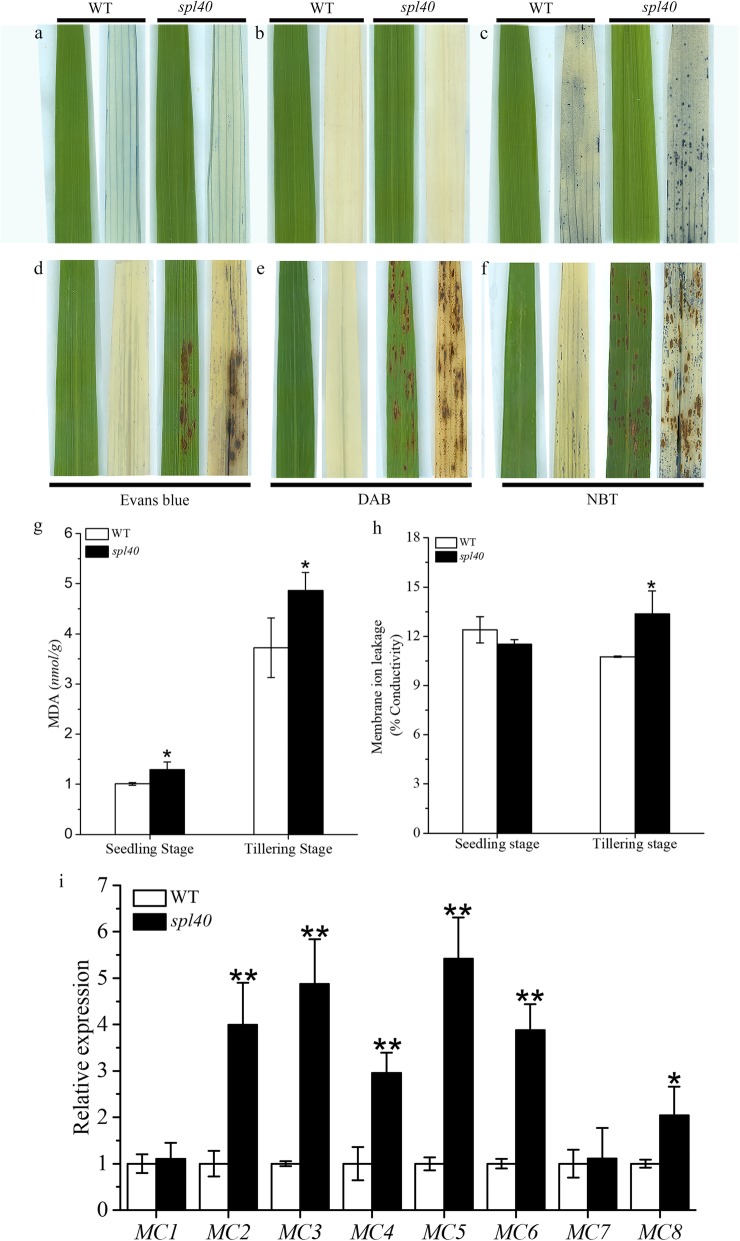
Fig. 3.
Histochemical staining of spl40 and WT. a and d Evans blue staining: a before lesion development. d after lesion development. b and e DAB staining: b before lesion development. e after lesion development. c and f NBT staining: c before lesion development. f after lesion development. g Malonaldehyde (MDA) content at seedling and tillering stage. h Membrane ion leakage rate at seedling and tillering stage. i Expression analysis of PCD-related genes at tillering stage. Values are means ± SD (n = 3); ** indicates significance at P ≤ 0.01 and * indicates significance at P ≤ 0.05 by Student’s t test

