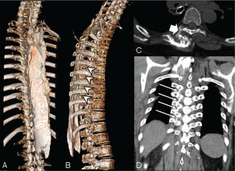
Figure 2.

Computed tomography scan and 3-dimensional reconstruction. (A) The contrast spread extensively in the cephalocaudal direction between the C4 and L1 vertebrae. (B) Arrow head indicate that the contrast spread to the costotransverse foramen at level of T6-T10. (C) Contrast spread to the thoracic paravertebral space (thick arrow). (D) On the coronal section, the contrast spread to the costotransverse ligament (arrows), which connects the rib and the transverse process.
