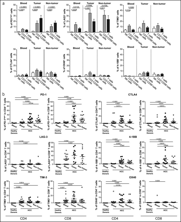
Figure 2.
Immune checkpoint distribution on lymphocyte subsets in advanced HCC at the baseline. (a) The percentage of immune checkpoint-positive cells among NK, CD3+CD56+ cells (NKT and CD3brightCD56+ T cells), and T cells in the blood (n = 21), tumor tissue (n = 16), and nontumoral tissue (n = 13). (b) The percentage of immune checkpoint-positive cells among CD4+ or CD8+ T cells in the blood (n = 21), tumor tissue (n = 16), and nontumoral tissue (n = 13). Each dot represents a patient. The nonparametric Kruskal-Wallis one-way analysis of variance was used for multiple comparisons. HCC, hepatocellular carcinoma.

