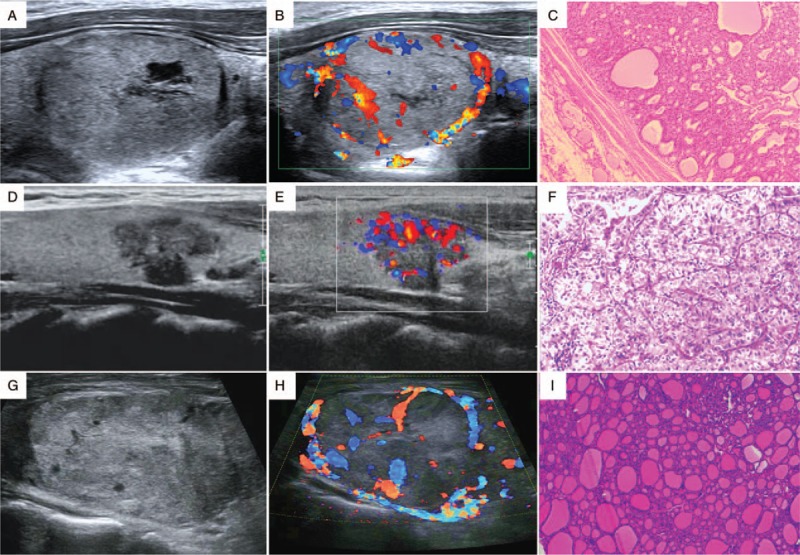
Figure 1.

Benign and suspicious ultrasound features and pathological findings (HE staining, original magnification ×200) of thyroid nodules. (A–C) A nodular goiter. The two-dimensional image, color Doppler image, and pathologic image are shown. (D–F) A papillary thyroid carcinoma. The ultrasound image shows a hypoechoic nodule with an irregular margin and microcalcification. (G) (H), and (I) A follicular carcinoma demonstrating a large isoechoic nodule without microcalcification. HE: Hematoxylin and eosin.
