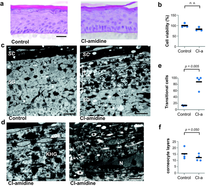
Figure 2. Increase of transitional cells and decreased number of corneocyte layers after Cl-amidine treatment.

Reconstructed human epidermis (RHEs) were treated with 800 μM Cl-amidine (Cl-a) for 48 h (n = 4). (a) Treated and control RHEs were stained with hematoxylineosin. Scale bar = 25 μm. (b) The cell viability was evaluated using the MTT assay. (c and d) RHEs were analyzed by transmission electron microscopy. Scale bars = 1 μm. At the transition between stratum granulosum (SG) and stratum corneum (SC), transitional cells (T) are frequently observed in treated RHEs. They are characterized by a marked cornified envelope (CE), diffuse keratohyalin granules (KHG) and cytoplasmic vesicles (asterisks). Sometimes the nucleus (N) is observed. (e and f) The frequency of transitional cells and the number of corneocyte layers were quantified. Mean values for each RHE are represented by blue dots and means of all experiments by black dashes.
