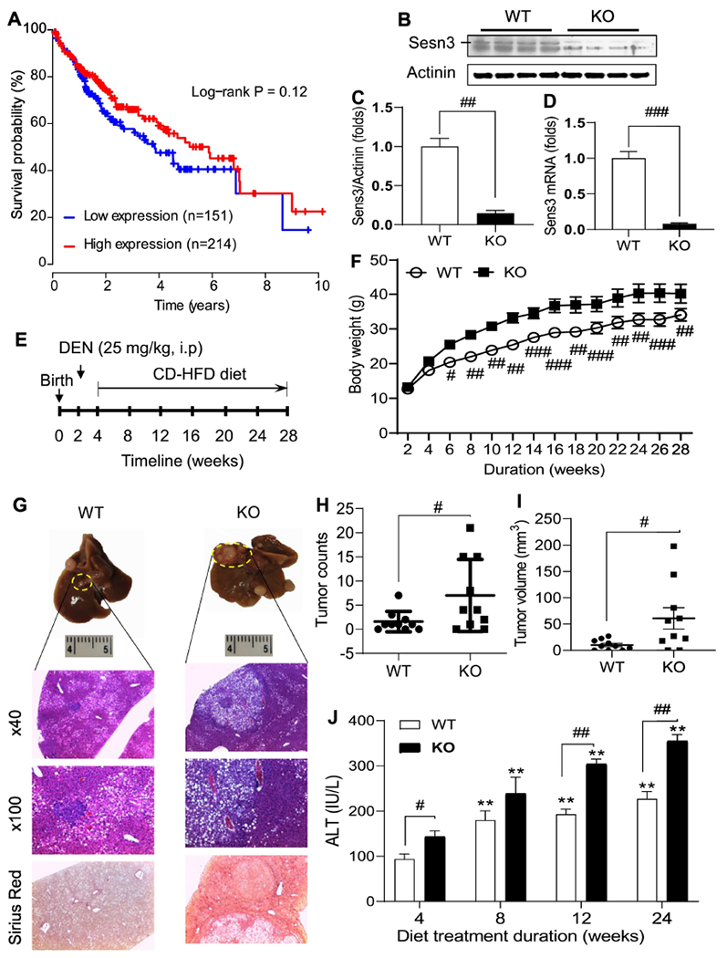
Fig 1. Sesn3 knockout mice are more susceptible to carcinogen-induced hepatocellular carcinoma.

Kaplan-Meier survival curve was generated using the TCGA liver cancer dataset (A). Sesn3 KO in liver tissues was confirmed by immunoblot analysis (B), protein quantification (C, n=4), and real-time PCR mRNA analysis (D, n=4). Experimental scheme for the pre-clinical HCC model in Sesn3 KO mice (E). Mouse body weights during the experimental period (F, n=10). Macroscopic and microscopic analysis of tumor-bearing liver tissue (G). Tumor nodule counts (H, n=10) and tumor volume (I, n=10) in the mouse hepatic tissue. Serum ALT measurements at multiple time points during the HCC development (J, n=10). Data are presented as mean ± SEM. #p < 0.05, ##p < 0.01, and ###p < 0.001 for KO vs. WT. **p < 0.05 for comparisons vs. 4 weeks in panel J.
