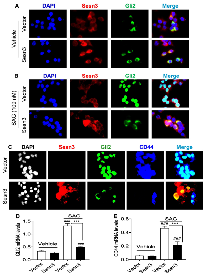
Fig 4. Sesn3 suppressed the Gli2 nuclear localization and CD44 gene expression.

Representative immunofluorescent images of Sesn3 and Gli2 after vector, Flag-Sesn3, and HA-Gli2 plasmid DNAs were transfected to Huh7 cells in the absence or presence of 100 nM SAG. Sesn3 and Gli2 were detected using anti-Sesn3 or anti-HA antibody followed by Alexa 594 and Alexa 488 secondary antibody, respectively (A, B). Vector or Flag-Sesn3 plus HA-Gli2 were transfected to Huh7 cells in the presence of 100 nM SAG. Sesn3, Gli2, and CD44 were detected using anti-Flag, anti-HA, and anti-CD44 antibody followed by Alexa 594, Alex 488, and Cy5 secondary antibody, respectively (C). Fluorescence images were captured using a Zeiss fluorescence microscope at x640 magnification. Real-time PCR analysis of endogenous GLI2 and CD44 mRNAs in vector or Sesn3 transfected Huh7 cells treated with vehicle or SAG (100 nM) for 24 hours (D, E). Data are expressed as mean ± SEM (n = 3). ###p < 0.001 for vehicle vs. SAG for vector or Sesn3 transfection, and ***p < 0.001 for vector vs. Sesn3 transfection after the SAG treatment.
