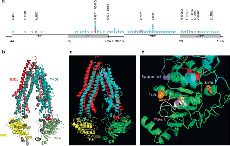
Figure 1. Molecular model of ABCB5.
An atomic model of ABCB5 was constructed based on the sequence alignment of full-length ABCB5 to mouse ABCB1 or P-glycoprotein, for which experimental structures are known. (a) Schematic of the ABCB5 protein topology with the conserved domains indicated as blocks, including transmembrane domains (TMD) and nucleotide binding domains (NBD). Somatic alterations are represented by arrowheads. A total of 71 different alterations were identified, and for clarity, only some amino acid changes were indicated. Underlined alterations were functionally assessed. Red triangles represent deleterious alterations, blue triangles represent missense mutations, the orange triangle represents splice site, and purple triangles represent silent mutations. (b) Ribbon diagram of the full-length ABCB5 model, with the N-terminal transmembrane domain, or TMD1 in red and the C-terminal TMD2 in cyan. The N-terminal NBD1 and the C-terminal NBD2 are shown in yellow and green, respectively. Residues where mutations were found are displayed as ball models. (c) Residues S830 in the TM8 is shown in magenta, and residues S1091 and S1184 in NBD2 are colored in orange. (d) Magnified view of NBD2 showing detailed structural environment in the vicinity of the two mutation sites in ball models in orange. The Walker A and B motifs are colored light orange and pink, respectively. The signature motif is in purple. The two intracellular helical motifs are shown in cyan and red, respectively.

