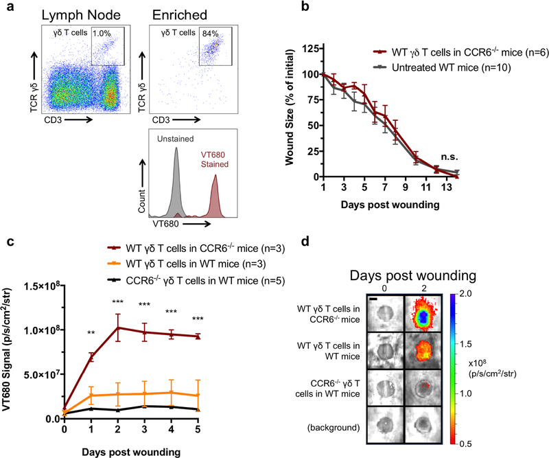
Figure 2: Adoptive transfer of γδ T cells restores wound healing in CCR6−/− mice.
(a) γδ T cells were enriched from the lymph nodes of WT and CCR6−/− mice and stained with the membrane dye VT680. 100,000 γδ T cells were transferred into mice via the tail vein. Mice were wounded the subsequent day, and (b) wound size was measured daily. (c and d) VT680 signal was measured for 5 days to measure γδ T cell trafficking. Data is presented as Mean ± SEM. * P **P ≤ 0.01 and ***P ≤ 0.001 comparing CCR6−/− mice receiving WT γδ T cells to other groups using a repeated measures two-way analysis of variance followed by Bonferroni’s post hoc test (b and c) Scale bar = 3 mm.

