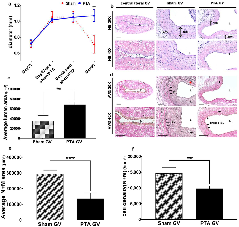
Figure 1: Diameter, lumen vessel area, and area of the neointima + media.
(a) By day 56, there is a significant increase in the average intraoperative diameter of the graft veins treated with PTA compared to sham controls (**P<0.01). B upper row are representative sections at 20X magnification and b lower row is at 40X magnification after Hematoxylin and eosin staining of the contralateral jugular control vein (contralateral CV), sham jugular graft vein (sham GV), and PTA treated jugular graft vein (PTA GV). (c) There is a significant increase in the average lumen vessel area of the PTA vessels compared to sham vessels (P<0.01). D upper row are representative sections at 20X magnification and d lower row is at 40X magnification after Verhoeff-van Gieson staining of the contralateral CV, sham GV, and PTA GV. For CV, there is no internal elastic lamina (IEL). In contrast, the IEL is intact in the sham GV. In the PTA GV, the IEL is broken. (e) There is a significant decrease in the average cell density of the neointima + media of the PTA vessels compared with sham vessels (**P<0.01). (f) There is a significant decrease in the average area of the neointima + media of the PTA vessels compared with sham vessels (***P<0.001). Each bar represents mean ± SEM of 6–7 animals. ADV, adventitia; L, lumen; N + M, neointima + media; IEL, internal elastic lamina; solid arrows, indicate the IEL; solid black star, neointima; solid red star indicates severe neointimal hyperplasia with loss of IEL. Scale bar is 50-μm.

