
Figure 1 (Continued).
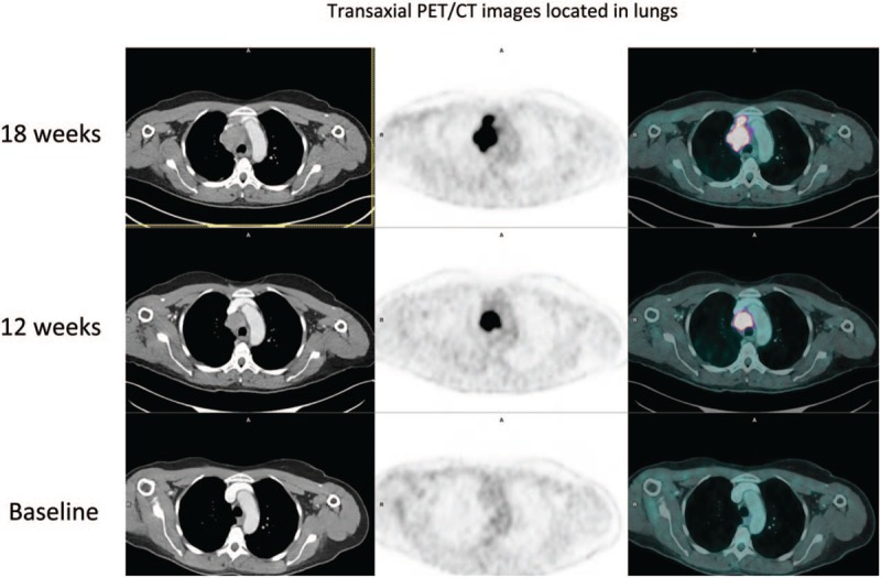
Maximum intensity projection (MIP)18F-FDG PET/CT images during the course of treatment in a 54-year-old woman with metastatic melanoma, A Baseline PET/CT (2 right lung nodules and 1 right hilar node), B Interim PET/CT (12 weeks) with progression non equivocal (PD according to RECIST 1.1 and PERCIST, iUPD according to iRECIST, no clinical benefit according to PECRIT), C Final PET/CT (18 weeks) with confirmation of progression disease (iUPD transformed in iCPD according to iRECIST).