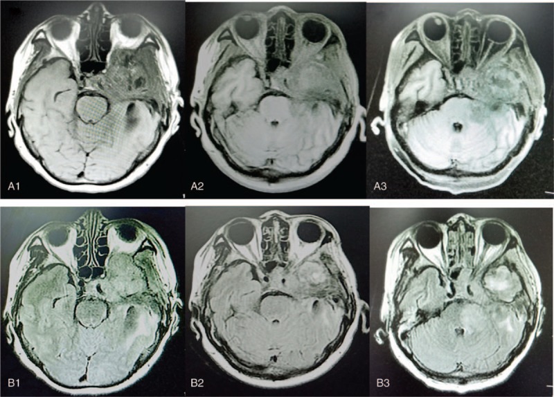
Figure 1.

MRI findings in patient presented with glioblastoma. A and B represent MRI T1 weighting and T2 weighting, respectively. 1, 2, and 3 represent before, 1 month after treatment, and 2 months after treatment of anlotinib, respectively. MRI = magnetic resonance imaging.
