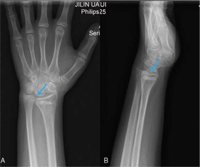
Figure 1.

(A) Frontal and (B) lateral radiographs of left wrist joint. An 11-year-old male patient displayed swelling and pain of the left wrist. According to the preoperative x-ray image, the distal part of the ulna is longer than that of radial (blue arrow).
