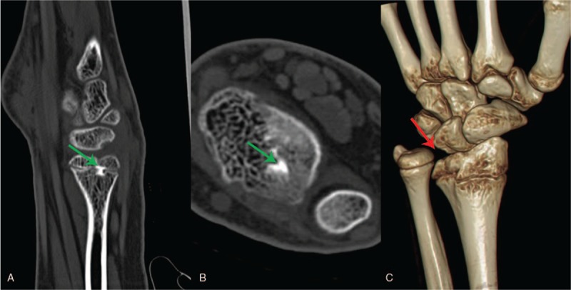
Figure 2.

Preoperative 3D CT of left wrist joint. It showed a strip-like high density area (green arrow) below the epiphyseal plate of the distal radius (A–B), and abnormal wrist anatomy (red arrow) (C). 3D = three dimensional, CT = computed tomography.
