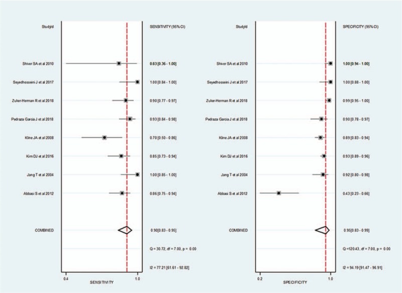
Figure 5.

Coupled forest plots of pooled sensitivity and specificity of 3-point point-of-care ultrasound for the diagnosis of deep vein thrombosis. Dots in squares represent sensitivity and specificity. Horizontal lines represent the 95% confidence interval (CI) for each included study. The combined estimate (“Summary”) is based on the random-effects model and is indicated using diamonds. Corresponding heterogeneities (I2) with 95% CIs are provided in the bottom right corners.
