
Figure 1.
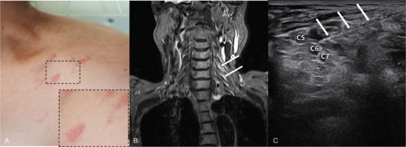
(A) The erythematous vesicular eruption is distributed around the left shoulder. (B) Brachial plexus MRI acquired 21 days after the onset of symptoms. Coronal T2-weighted short tau inversion recovery images demonstrate mild swelling and increased signals in the left brachial plexus especially in the C5 and C6 dorsal root ganglia and roots (white arrow) (C) Ultrasound-guided PDRN injection (white arrow) to the left C5 and C6 root. MRI = magnetic resonance images, PDRN = polydeoxyribonucleotide.