Figure 2.

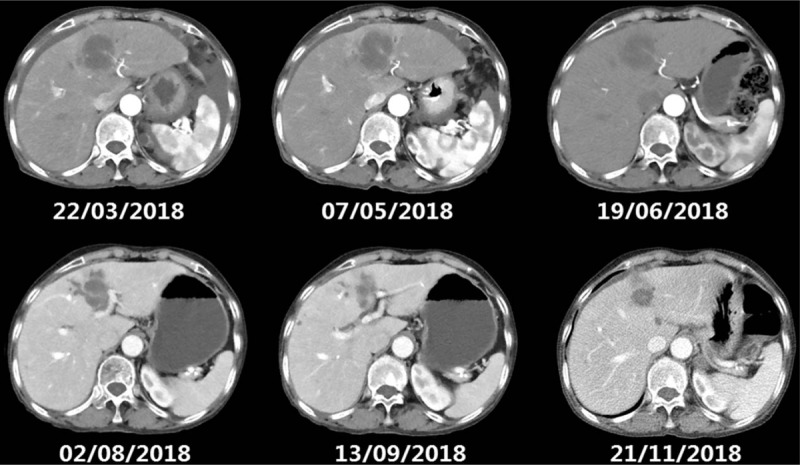
The following CT scan shows mass lesion in the left lobe of liver remained stable (from February to July) and then reduced. CT = computed tomography.

The following CT scan shows mass lesion in the left lobe of liver remained stable (from February to July) and then reduced. CT = computed tomography.