
Figure 1.
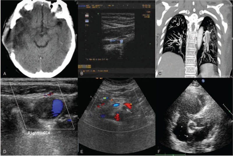
Imaging examinations. Cranial computed tomography (CT) indicated changes after the right temporal decompressive craniectomy (A). Vascular ultrasound revealed solid low echoes at the distal end of the right subclavian artery (B). computed tomography pulmonary arteriography (CTPA) showed wide embolism at the distal end of the bilateral main pulmonary arteries (C). A vascular ultrasound of the neck revealed solid low echoes at the right internal carotid artery (D). The mesenteric arteriovenous ultrasound showed that, the sonolucency of the main trunk and partial branches was poor with no blood flow filling (as indicated by the arrows) (E). Echocardiography indicated the color trans-septal blood flow signals were explored at the oval foramen (as indicated by the arrows) (F).