
Figure 2.
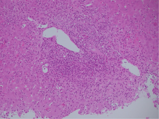
An image of a liver biopsy specimen (Hematoxylin and Eosin staining, original magnification ×200). The presence of interface hepatitis with dense portal lymphocyte infiltrate disrupting the limiting plate is shown.
Official websites use .gov
A
.gov website belongs to an official
government organization in the United States.
Secure .gov websites use HTTPS
A lock (
) or https:// means you've safely
connected to the .gov website. Share sensitive
information only on official, secure websites.
An image of a liver biopsy specimen (Hematoxylin and Eosin staining, original magnification ×200). The presence of interface hepatitis with dense portal lymphocyte infiltrate disrupting the limiting plate is shown.