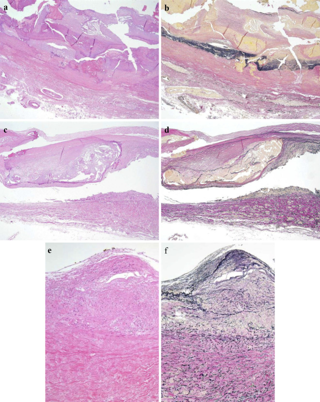
Figure 3.
Histopathological findings of the affected aorta. (a, b) Thoracoabdominal aorta, (c, d) common iliac artery, (e, f) right renal artery. Fibrous thickening and calcification were demonstrated in the intima, with rupture and disappearance of the medial elastic fibers. Fibrous thickening of the adventitia and feeding vessels in the affected aorta was found. Inflammatory cell infiltration in the adventitia was very slight. (a, c, d) Hematoxylin and Eosin staining, (b, d, f) Elastica van Gieson (EVG) stain. (a-d) ×20, (e-f) ×100

