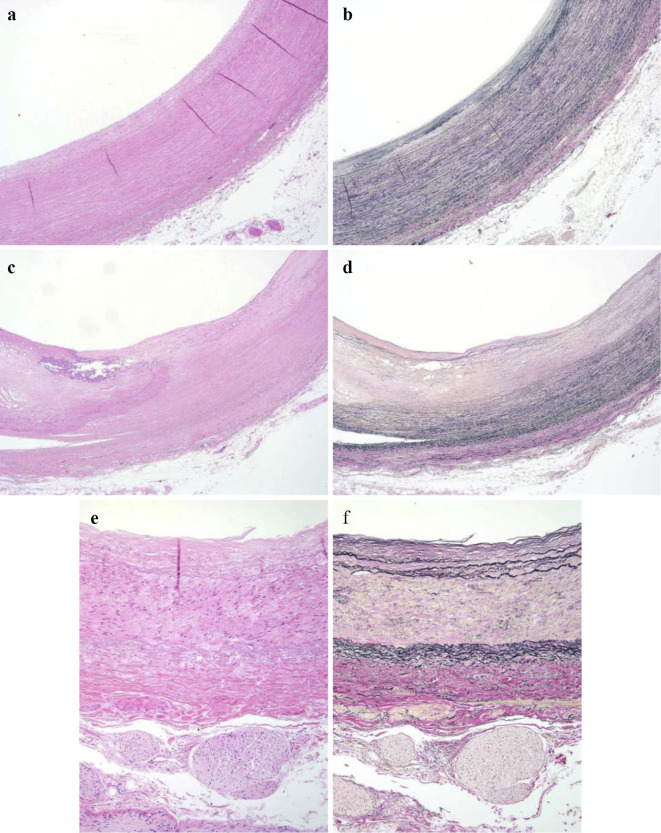
Figure 4.
Histopathological findings of unaffected arteries. (a, b) Brachiocephalic artery, (c, d) left subclavian artery, (e, f) left renal artery. The medial elastic fibers were preserved with only slight atherosclerosis. (a, c, e) Hematoxylin and Eosin staining. (b, d, f) EVG stain. (a-d) ×20, (e-f) ×100

