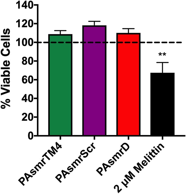
FIG 4.

Propidium iodide uptake as a measure of membrane disruption in response to peptide addition. Overnight cultures of E. coli cells expressing PAsmr were resuspended in minimal medium A, representing viable cells. Cells were diluted to an OD600 of 0.1, and a standard curve was generated using the viable cells and cells 100% disrupted by 70% isopropanol. Diluted cells in minimal medium A were then incubated with 8 μM peptide or 2 μM melittin, followed by the addition of 5 μM PI. Fluorescence was measured, and using the equation of the standard curve, the relative amounts of viable cells were calculated. Error bars show standard deviations (n ≥ 3). **, P > 0.001 for comparison of melittin treatment to PAsmrTM4 and PAsmrScr treatments; no significant difference was noted among peptides.
