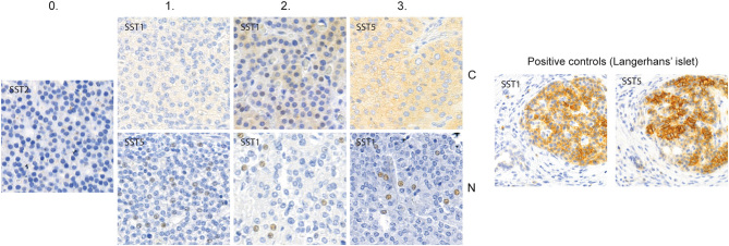
Figure 1.
Examples of the staining intensities in parathyroid tumor tissue for the scoring system used in our study for cytoplasmic and nuclear expression. Membrane expression was weak and the intensity was not sufficiently diverse to make a similar comparison. Left: negative staining (0). Subsequent: weak cytoplasmic and nuclear staining (1), strong cytoplasmic and nuclear staining (2) and very strong cytoplasmic and nuclear staining (3). On the right are pancreatic islets stained with SST1 and SST5 presenting cytoplasmic and membrane positivity, functioning as positive control. Nuclear positivity is not found in the control stainings.

 This work is licensed under a