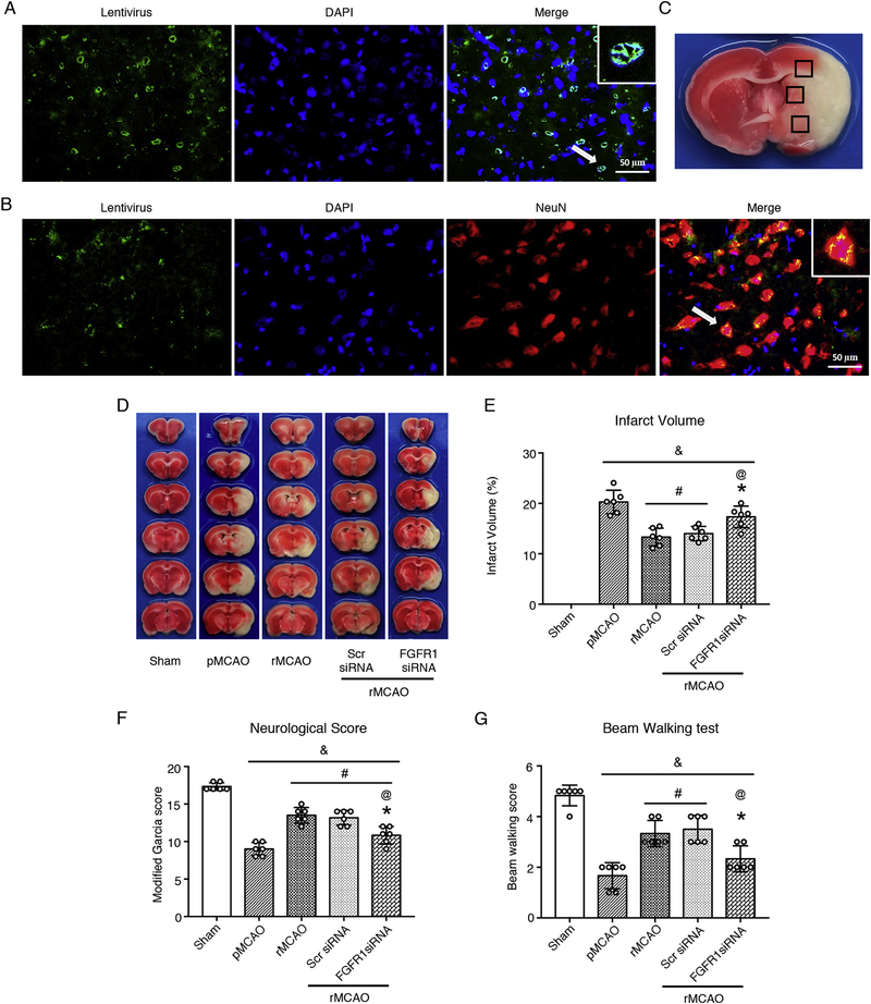
Fig. 6.
FGFR1 siRNA weakened the effects of recanalization on neurological outcome at 10 d after MCAO. (A–B) Representative microphotographs of FGFR1 siRNA lentivirus (GFP, Green) transduced neurons (NeuN, Red) at 4 d after intracerebroventricular injection, A was observed after rat sacrifice at once, B was observed at 24h after sacrifice with NeuN staining. DAPI (Blue) marked Nuclei. Scale bar = 50 μm. (C) Samples were obtained from the ischemic penumbra. (D) Representative images of TTC stained brain slices. TTC staining was only used to calculate infarct volume; (E) Quantified infarct volume; (F) Modified Garcia score, and (G) Beam walking score, & p < 0.05, vs sham, # p < 0.05, vs pMCAO, * p < 0.05, vs rMCAO, @ p < 0.05, vs Scr siRNA, n = 6 per group, One-way ANOVA-Tukey. NeuN, neuronal nuclei; DAPI, 4’,6-diamidino-2-phenylindole; pMCAO, permanent middle cerebral artery occlusion; rMCAO, recanalization at 3 d after MCAO; Scr siRNA, scrambled siRNA; FGFR1, fibroblast growth factor receptor 1.

