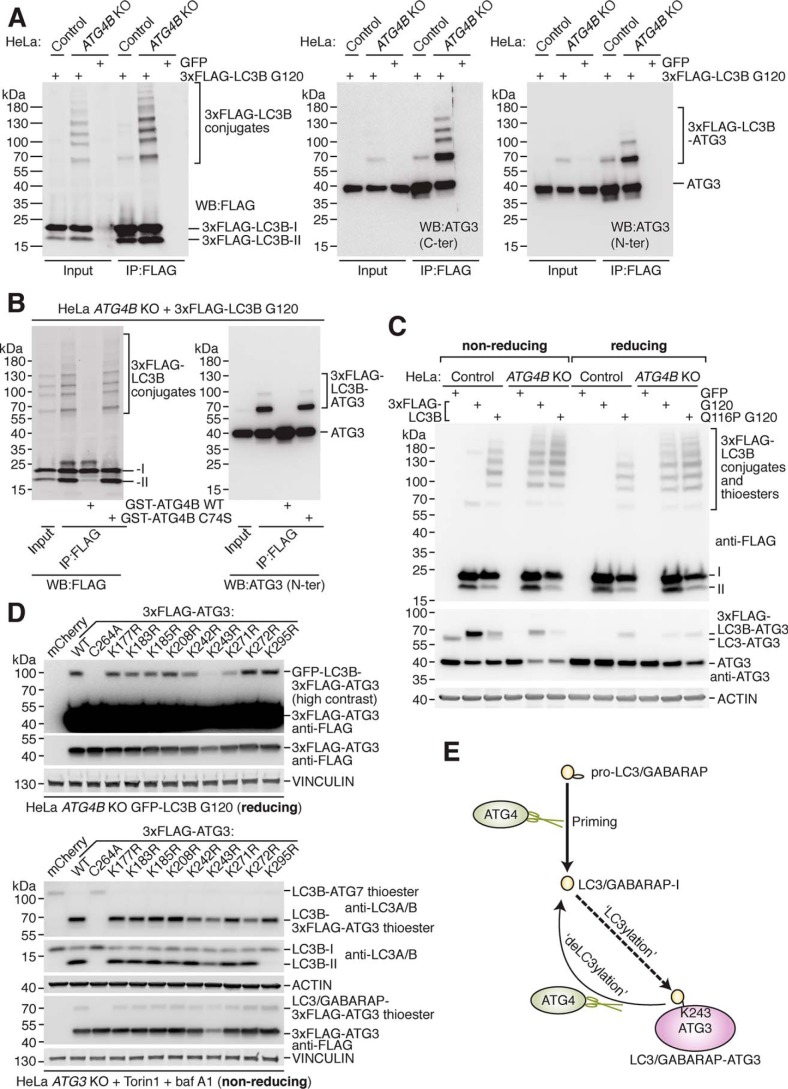
Figure 3.
ATG3 protein is a target of LC3/GABARAP conjugation and ATG4-mediated deconjugation. A, enrichment of transiently expressed 3xFLAG–LC3B G120 from HeLa control and ATG4B KO cells by IP with anti-FLAG antibody prior to Western blotting with anti-FLAG antibody (left) and anti-ATG3 antibodies (middle, right panel). Cells were transfected with GFP as a negative control. B, 3xFLAG–LC3B G120 expressed in HeLa ATG4B KO cells was enriched by IP with anti-FLAG antibody prior to treatment with 0.02 mg/ml recombinant GST–ATG4B WT or C74S at 37 °C for 1 h and Western blotting using anti-FLAG and anti-ATG3 N-ter antibody. C, HeLa control and ATG4B KO cells were transfected with 3xFLAG–LC3B G120, 3xFLAG–LC3B Q116P G120, or GFP as a negative control prior to harvesting. Western blotting was performed to detect 3xFLAG–LC3B and ATG3 using the same lysates diluted with either nonreducing (β-mercaptoethanol excluded) or reducing (standard recipe) sample buffer. Thioester-linked species are detected under nonreducing conditions only. D, Western blotting of HeLa ATG4B KO GFP–LC3B G120 cells transiently transfected with 3xFLAG–ATG3 WT and point mutant constructs. Samples were lysed in buffer lacking NEM and processed under reducing conditions (upper panel). The same constructs were used to rescue HeLa ATG3 KO cells treated for 3 h with 250 nm Torin1 and 10 nm baf A1 and assessed by Western blotting under nonreducing conditions (lower panel). E, schematic of human ATG4 protease cellular function in protein deconjugation (deLC3ylation), which counteracts covalent attachment of LC3/GABARAP (LC3ylation) to ATG3 protein at residue Lys-243.

