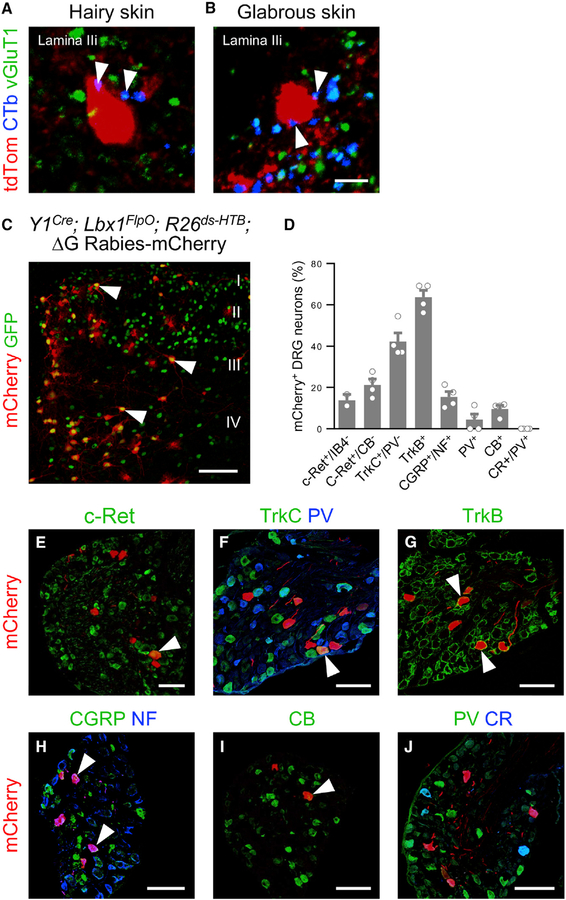
Figure 2. Y1Cre Neurons Receive Extensive LTMR Input.
(A and B) Examples of Y1Cre neurons in lamina IIi from lumbarspinal cord sectionsof P42 Y1Cre; Ai14lsl-tdTom mice injected with CTb into: the hairy skin of the thigh (A) and the glabrous skin of the hindpaw (B). Immunolabeled CTb+ contacts (blue) displayed vGluT1 immunoreactivity (green, arrowheads).
(C) Section through the lumbar dorsal horn of a P10 Y1Cre; Lbx1FlpO; R26ds-HTB mouse injected with EnvA G-deleted rabies-mCherry virus. Arrowheads indicate infected Y1Cre neurons. mCherry+/GFP— cells represent transsynaptically labeled presynaptic neurons.
(D) Summary of antibody-labeled myelinated sensory afferent subtypes that are presynaptic to the Y1Cre neurons, expressed as a percentage of mCherry+ neurons (n = 4 mice).
(E–J) Sections from P10 Y1Cre; Lbx1FlpO; R26ds-HTB lumbar DRGs showing presynaptically labeled sensory neurons(red) thatexpress c-Ret (E), TrkC but not parvalbumin(PV; F), TrkB(G), calcitonin gene-related peptide (CGRP) and neurofilament 200 (NF; H), and calbindin (CB; I), but not PV or calretinin (CR; J). Arrowheads indicate co-labeled sensory afferents. CB, calbindin; CR, calretinin; NF, neurofilament; PV, parvalbumin. Scale bars: 5 μm (B) and 100 μm (C and E–J). Data: mean ± SEM. See also Figure S4.

