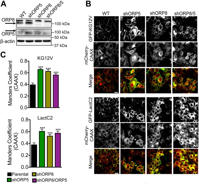
Figure 3. Knockdown of ORP5 or ORP8 mislocalizes KRAS and PtdSer from the PM.
(A) shRNA knockdown of ORP5 and ORP8 separately and simultaneously in MCF-7 breast cancer cells was validated by Western blotting, and β-actin levels were used as loading controls. (B) Parental (WT) and knockdown cells were transiently transfected with GFP-KRASG12V and mCherryCAAX (an endomembrane marker) or GFP-LactC2 and mCherryCAAX and imaged in a confocal microscope. Representative images are shown. (C) The extent of KRAS and LactC2 mislocalization was quantified using Manders coefficient, which measures the extent of colocalization/overlap of GFP and mCherry signals. Significant differences were evaluated using t tests (±SEM, n ≥ 6) (*P < 0.05, **P < 0.01, ***P < 0.001); scale bar 20 μm.

