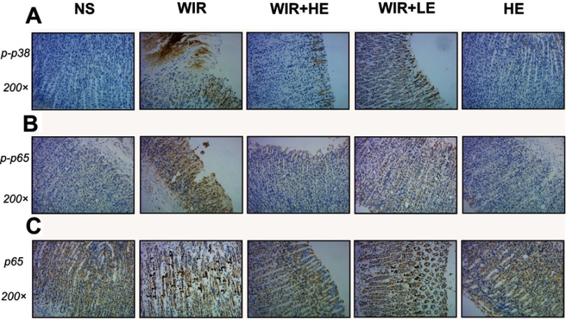
Figure 8.
Representative pictures showing the immunohistochemical analysis of p-p38, p-p65 and NF-κB p65 in sections of gastric mucosa obtained from rats.
Notes: (A and B) The immunohistochemical staining of p-p38 and p-p65 in the gastric tissues (magnification, 200×) in different groups, brown staining denotes positive expression. (C) The immunohistochemical detection of the nuclear translocation of p65 subunit in gastric tissues, ▲ labels epithelial cells with hyperchromatic area around nucleus (magnification, 200×) in different groups.
Abbreviations: p-p38, phosphorylated p38; p-p65, phosphorylated p65; NF-κB, nuclear factor kappa B.

