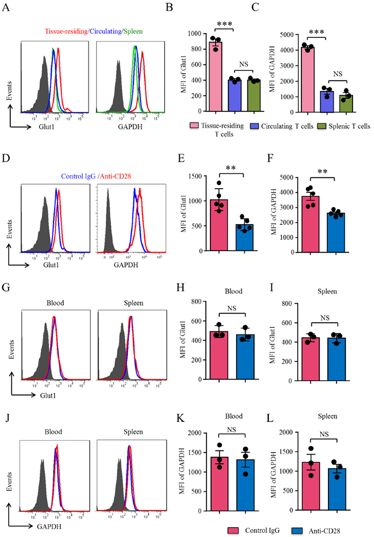
Figure 4. CD28 signaling regulates glucose utilization in tissue-residing CD4 T-cells.

(A-C) Vasculitis was induced as in Figure 1. Spleens and arteries were harvested and digested. Protein expression of the glucose transporter Glut1 and the glycolytic enzyme GAPDH was analyzed by flow cytometry in splenic, peripheral blood and artery-infiltrating CD4+ T-cells. Representative histograms and mean fluorescence intensities (MFIs) from 3 experiments (t test). (D-F) Human artery-NSG chimeras were treated with anti-CD28 dAb or control Ab as in Figure 1. Cells were isolated from the explanted arteries, spleens and blood and Glut1 and GAPDH expression on CD4 T-cells was determined by multi-color flow cytometry. Representative histograms and MFIs from 5 experiments in E and F and 3 experiments in G to L (t test). All FACS plots were gated on CD45+CD4+ cells. Data are mean±SEM. **p<0.01, ***p<0.001. ns: not significant. GAPDH: Glyceraldehyde-3-phosphate dehydrogenase; Glut1: Glucose transporter 1.
