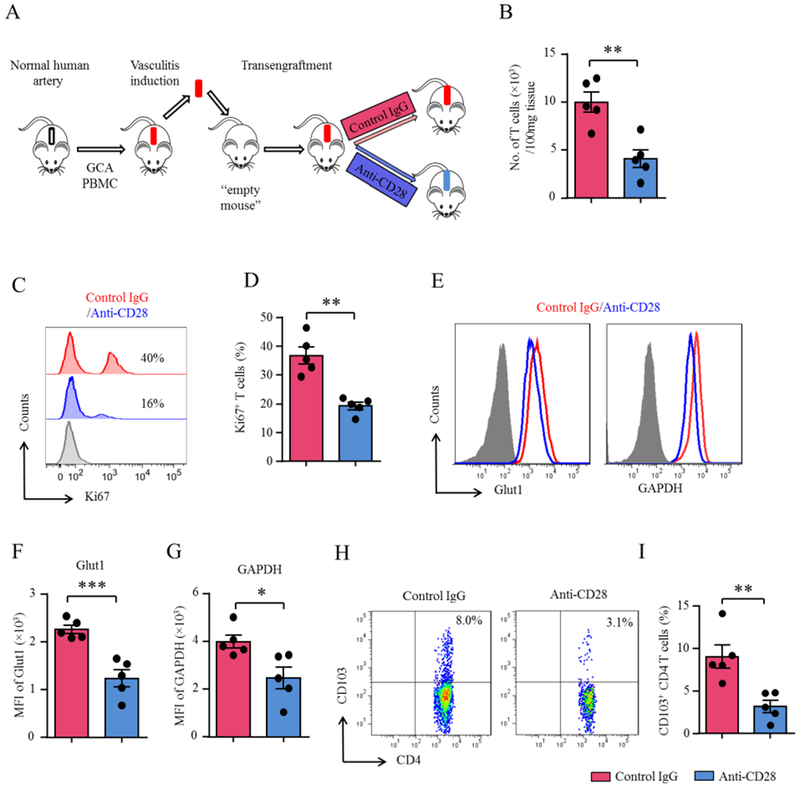
Figure 5. Tissue-resident memory T cells depend on CD28 signaling.

(A) Scheme of the mouse experiments. (B-I) Vasculitis was induced in human arteries as in Figure 1. To analyze cells contained within the vasculitic lesions, inflamed arteries were trans-engrafted into an “empty” NSG mouse, which were treated with anti-CD28 dAb or control Ab (5mg/kg, 4x/week). Single cell suspensions were prepared from explanted arteries. (B) Enumeration of tissue-residing CD4+ T-cells (t test). (C-D) Quantification of proliferating T-cells in the vasculitic lesions by Ki-67 flow cytometry. Representative histograms and percentages of Ki-67+ CD4+ T-cells (t test). (E-G) FACS analysis of Glut1 and GAPDH expression on tissue-residing CD4+ T-cells. Representative histograms and fluorescence intensity (MFIs) from 5 experiments (t test). (H-I) CD103+ tissue-resident memory CD4 T-cells analyzed by multi-color flow cytometry. Representative FACS plots and results from 5 experiments (t test). All FACS plots were gated on CD45+CD4+ cells. All data are mean ± SEM. *p<0.05, **p<0.01, ***p<0.001. GAPDH: Glyceraldehyde-3-phosphate dehydrogenase; Glut1: Glucose transporter 1.
