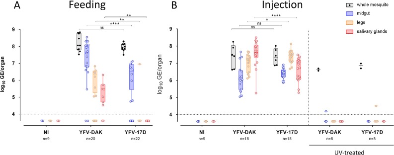
Fig 4. YFV-17D and YFV-DAK replicate in secondary organs when inoculated intra-thoracically.
(A) Mosquitoes were orally infected via a blood meal containing 4.107 PFU/mL of YFV-DAK, YFV-17D or no virus. Alternatively (B), mosquitoes were inoculated intra-thoracically with 2.5x10⁴ PFU of YFV-17D or YFV-DAK or with the same amount of UV-treated viruses. The relative amounts of organ-associated viral RNA were determined by RT-qPCR analysis 10 days after infection and are expressed as genome equivalents (GE) per organ. Several whole mosquitoes were also analyzed the day of the feeding or injection to ensure that a similar amount of viral particles of both viral strains were delivered in mosquitoes (black boxes). The number of organs (n) analyzed is indicated. The dashed lines indicate the limit of detection. Three independent experiments were performed with untreated viruses. Control experiments with UV-treated viruses were performed once. Statistical analyses were performed using a Mann-Whitney test (* p < 0.05; ** p < 0.01; *** p < 0.001; **** p < 0.0001).

