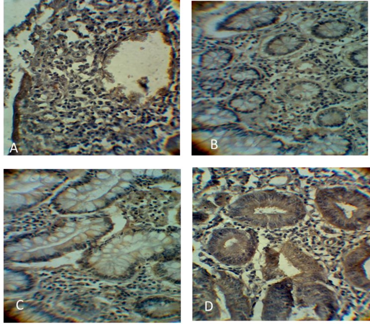
Fig. 2.
Immunohistochemistry staining of ABCG2 protein in both benign and cancererous colon tissues. A: low ABCG2 protein expression in benign tissues; B: stage I colon cancer showing positive ABCG2 in cytoplasm and cell membrane; C: stage II colon cancer with increased ABCG2 expression level; D: stage III colon cancer with high ABCG2 expression in tissues. All images were magnified at 400X

