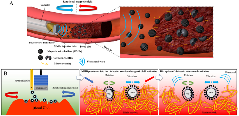
Figure 11:

Schematic view of sonothrombolysis with magnetic microbubbles under a rotational magnetic field. (A) MMBs are injected through the catheter and trapped near the clot region under the rotational magnetic field. (B) The hypothesis of sonothrombolysis with magnetic microbubbles under a rotational magnetic field. The MMBs can penetrate the clot surface and bind to the fibrin network of the clot under ultrasound exposure. When the rotational magnetic field is activated at this point, the MMBs start to rotate and vibrate, and the resulting shear force will cause the mechanical injury to the fibrin network. Then the MMBs can be activated by the ultrasound exposure and induce the cavitation effects, leading to the disruption of the clot.
