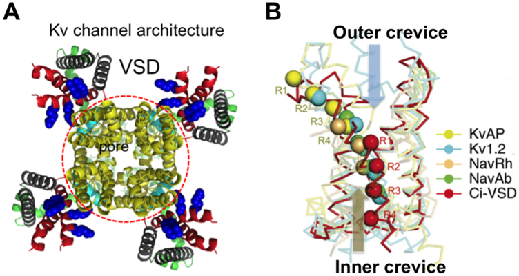
Figure 2: Architecture of a Kv channel and variations in VSD structures.
A) KvAP model after the Kv2.1 structure showing a central pore domain (red circle) and four VSDs. B) Five VSD structures in alignment. Arginine residues on S4 are aligned across the gating pore. Outer (upper) and inner (lower) crevices separated by a hydrophobic gasket (red). In a full resting (down) state, all four Arg residues move to the inner crevice, and drive the pore domain into the closed state [1, 2]. Panel B was modified from ref 1.

