Figure 1.
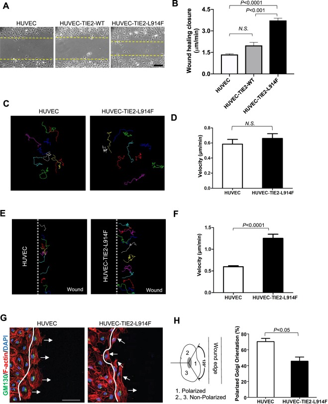
Constitutive active mutant TIE2 increases EC migration in response to wound healing. (A) Phase contrast pictures of the wound migration assay of HUVEC (human umbilical vein endothelial cells), HUVEC-TIE2-WT (wild-type) and HUVEC-TIE-L914F, 5 hours after scratching. Dashed lines indicate the wound closure front of migrating EC. Scale bar: 100 μm. (B) Quantification of wound healing closure speed (n = 3 independent experiments). (C) Analysis of single cell trajectories in non-confluent conditions over a 2-hour time course. (D) The cell velocity of 10–12 cells in a non-confluent monolayer was quantified in HUVEC and HUVEC-TIE2-L914F cells (n = 4 independent experiments). (E) Analysis of single cell trajectories at the migrating front of a wound healing assay. (F) The cell velocity of 10–12 cells at the migration front of a wound healing assay was quantified in HUVEC and HUVEC-TIE2-L914F cells (n = 4 independent experiments). (G) Immunofluorescence staining for GM130 (green), Phalloidin (F-actin) (red) and DAPI (blue). Scale bar: 50 μm. (H) Quantification of % of cells with polarized Golgi orientation on the moving front of the wound, two hours after scratching (n = 3 independent experiments).

