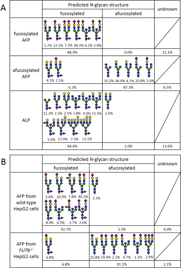
Figure 3.
Schematic representation of the glycoforms in the proteins used in this study. (A) Schematic representation of the glycoforms present in fucosylated AFP, afucosylated AFP, and ALP. (B) Schematic representation of the glycoforms present in AFP produced by wild-type and FUT8−/− HepG2 cells. Blue squares, green circles, yellow circles, yellow squares, magenta diamonds, and red triangles correspond to N-acetylglucosamine, mannose, galactose, N-acetylgalactosamine, sialic acid, and fucose, respectively.

