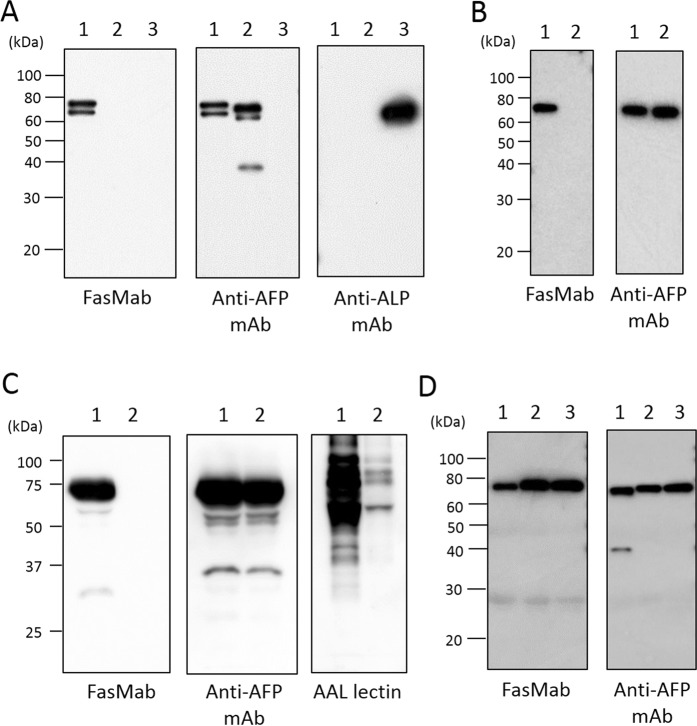
Figure 4.
Western blot analyses. (A) Western blot analyses for purified fucosylated AFP, afucosylated AFP, and ALP. Lane 1: fucosylated AFP, Lane 2: afucosylated AFP, Lane 3: ALP. (B) Western blot analyses for AFP purified from conditioned media of wild-type and FUT8-deficient HepG2 cells. Lane 1: AFP from wild-type HepG2 cells, Lane 2: AFP from FUT8-deficient HepG2 cells. (C) Western blot and AAL blot analyses for conditioned media of wild-type and FUT8-deficient HepG2 cells. Lane 1: Supernatant of wild-type HepG2 cells, Lane 2: Supernatant of FUT8-deficient HepG2 cells. (D) Western blot analyses for AFP following co-precipitation with an anti-AFP polyclonal antibody from human serum. Lane 1: sample 1, Lane 2: sample 2, Lane 3: sample 3. All images are detected individually, and cropped from different blots. Full-length blots of (A–D) are presented in Supplementary Figs 1–4, respectively.

