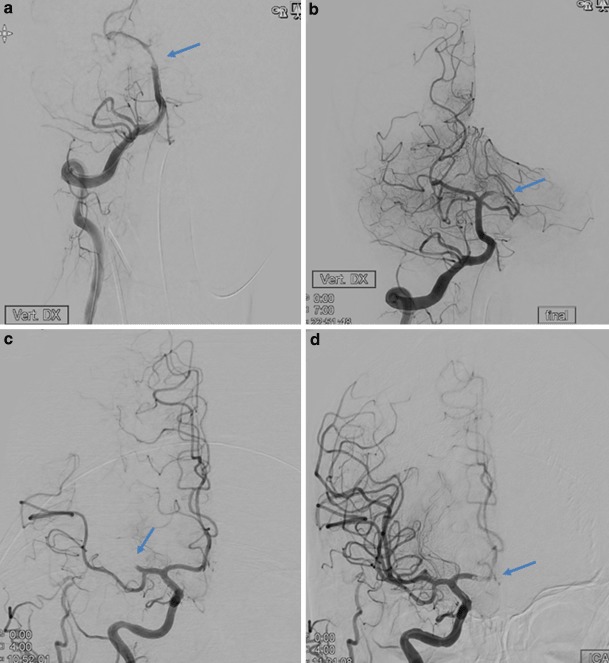
Fig. 1.

Computed tomography angiography. a and b Distal embolization in the same vascular territory: a angiographic run showing basilar artery occlusion (arrow showing location of occlusion), b after thrombectomy there is a left posterior cerebral artery thrombus (arrow showing new occlusion). c and d Distal emboli in a previously unaffected territory. c Right middle cerebral artery occlusion (arrow showing MCA occlusion). d After thrombectomy there is now an anterior cerebral artery thrombus (arrow showing new occlusion in the ACA)
