FIGURE 4.
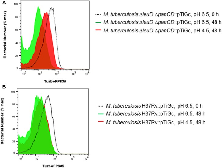
Fluorescence dilution demonstrates reduced replication of M. tuberculosisΔleuDΔpanCD under acid stress in comparison to M. tuberculosis H37Rv. M. tuberculosis H37Rv and M. tuberculosisΔleuDΔpanCD containing pTiGc was cultured in the presence of 4 mM theophylline (Theo), before removal of theophylline and exposure to pH 4.5 and pH 6.5 for 48 h prior to analyses by flow cytometry. Flow cytometry histograms demonstrate increased TurboFP635 fluorescence intensity in acidic media (pH 4.5, red), compared to normal media (pH 6.5, green) in M. tuberculosisΔleuDΔpanCD (A) after 48 h. In contrast, M. tuberculosis H37Rv demonstrated similar, reduced, TurboFP635 fluorescence intensity after 48 h in acidic media (pH 4.5, red) and control media (pH 6.5, green) (B). Representative examples of three independent biological repeats are shown.

