Figure 3.

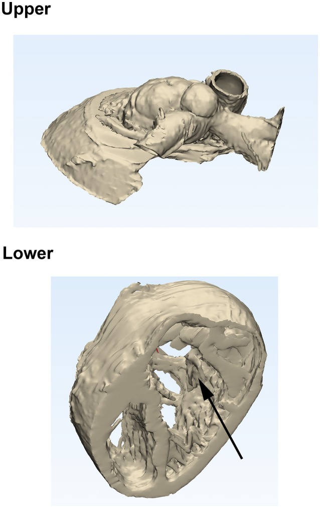
Model produced from later CT scan shortly before surgery. Screenshot from Materialize 3-Matic showing how the model would be cut (Upper and Lower portions), and demonstrating the tricuspid valve (arrow).

Model produced from later CT scan shortly before surgery. Screenshot from Materialize 3-Matic showing how the model would be cut (Upper and Lower portions), and demonstrating the tricuspid valve (arrow).