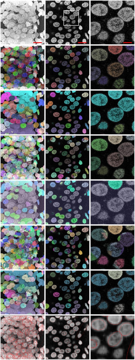
Figure 6.

Comparison of segmentation results with different other methods: (A) original image, (B) proposed method, (C) Ilastik, (D) gradient flow tracking, (E) MorphoLibJ, (F) Farsight, (G) classical watershed, (H) method based on the curvatures of the iso-intensity surfaces. Left column: z-projection, middle column: z-section, right column: magnification. Scale bars: 7 μm—left, middle, 2.3 μm—right. Segmented nuclei were randomly colorized for illustrative purposes (A–G), for (H) contours of ellipsoids representing the segmented results are shown.
