Figure 6.
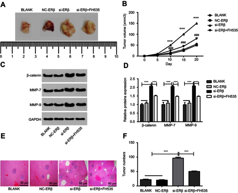
The growth and metastasis of OS tumors in mice. (A) Subcutaneous tumors under naked eye; (B) subcutaneous tumor volumes at different time points; (C) protein brands of Western blot; (D) relative expression of β-catenin, MMP-7 and MMP-9 at protein level (Western blot); (E) metastatic tumors in the liver tissues of mice under microscope (HE staining) (bar =50 μm, ×40); (F) the number of metastatic tumors. si-ERβ, U2-OS cells transfected with siRNA-ERβ for 48 hrs; NC-ERβ, U2-OS cells transfected with siRNA-negative control-ERβ for 48 hrs; si-ERβ + FH535, U2-OS cells transfected with siRNA-ERβ and treated with 20 μmol/L FH535 for 48 hrs; blank, U2-OS cells without transfection and treatment. ***P<0.001 vs NC-ERβ and blank; ###P<0.001 vs si-ERβ.
Abbreviations: ERβ, estrogen receptor β; OS, ostemsarcoma; GAPDH, glyceraldehyde-3-phosphate dehydrogenase; MMP, matrix metalloproteinase; HE, hematoxylin-eosin; NC, negative control; si, small interfering RNA.

