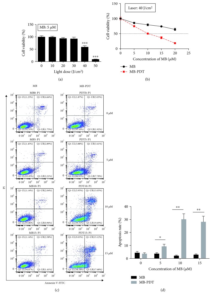
Figure 1.
MB-PDT induced apoptosis in THP-1 macrophages. (a) The survival rates of THP-1 macrophages following different laser irradiation doses (ranging from 0 to 50 J/cm2) with 5 μM MB. (b) The survival rates of THP-1 macrophages following 40 J/cm2 laser irradiation with different MB concentrations (0–20 μM). The results are expressed as the percentage of viable cells compared with medium alone-treated controls. (c) The apoptosis rate of THP-1 macrophages following photosensitization experiments (MB ranging from 0 to 15 μM with or without 40 J/cm2 irradiation) was assessed at 6 h posttreatment by Annexin V-FITC/PI binding and measured by flow cytometry analysis. The numbers indicate the percentage of cells in each quadrant. Specific statistical analyses are presented in (d). The results are expressed as the percentage of apoptotic cells (mean ± SD, n = 3). ∗P < 0.05, ∗∗P < 0.01, and ∗∗∗P < 0.001.

