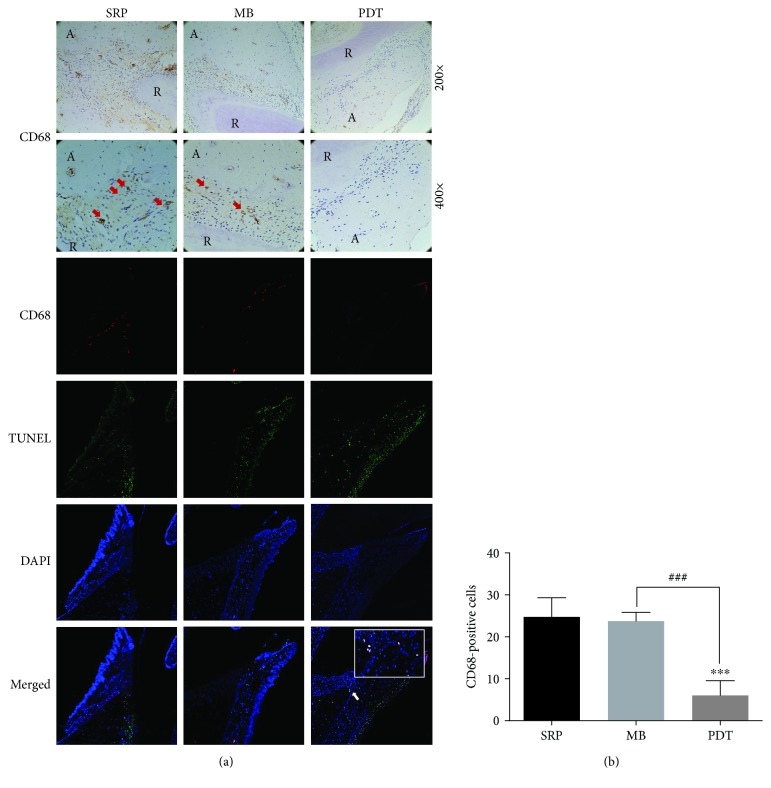
Figure 4.
Immunohistochemical and TUNEL staining of periodontal CD68 macrophages in rats. (a) Representative images of coronal sections of maxillary first molars from SD rats inoculated intraorally with Pg ATCC 33277 and Fn ATCC 25586 and probed with anti-rat CD68 mAb. Macrophages (CD68+ cells) in the periodontium are indicated by red arrows (original magnification 200x/400x). Apoptotic macrophages (CD68+ TUNEL+ cells) in the periodontium are indicated by white arrows (original magnification 100x). (b) CD68-positive cells are expressed as the mean ± SD (n = 8). ∗P < 0.05, ∗∗P < 0.01, and ∗∗∗P < 0.001. Abbreviations: R: root; A: alveolar bone.

