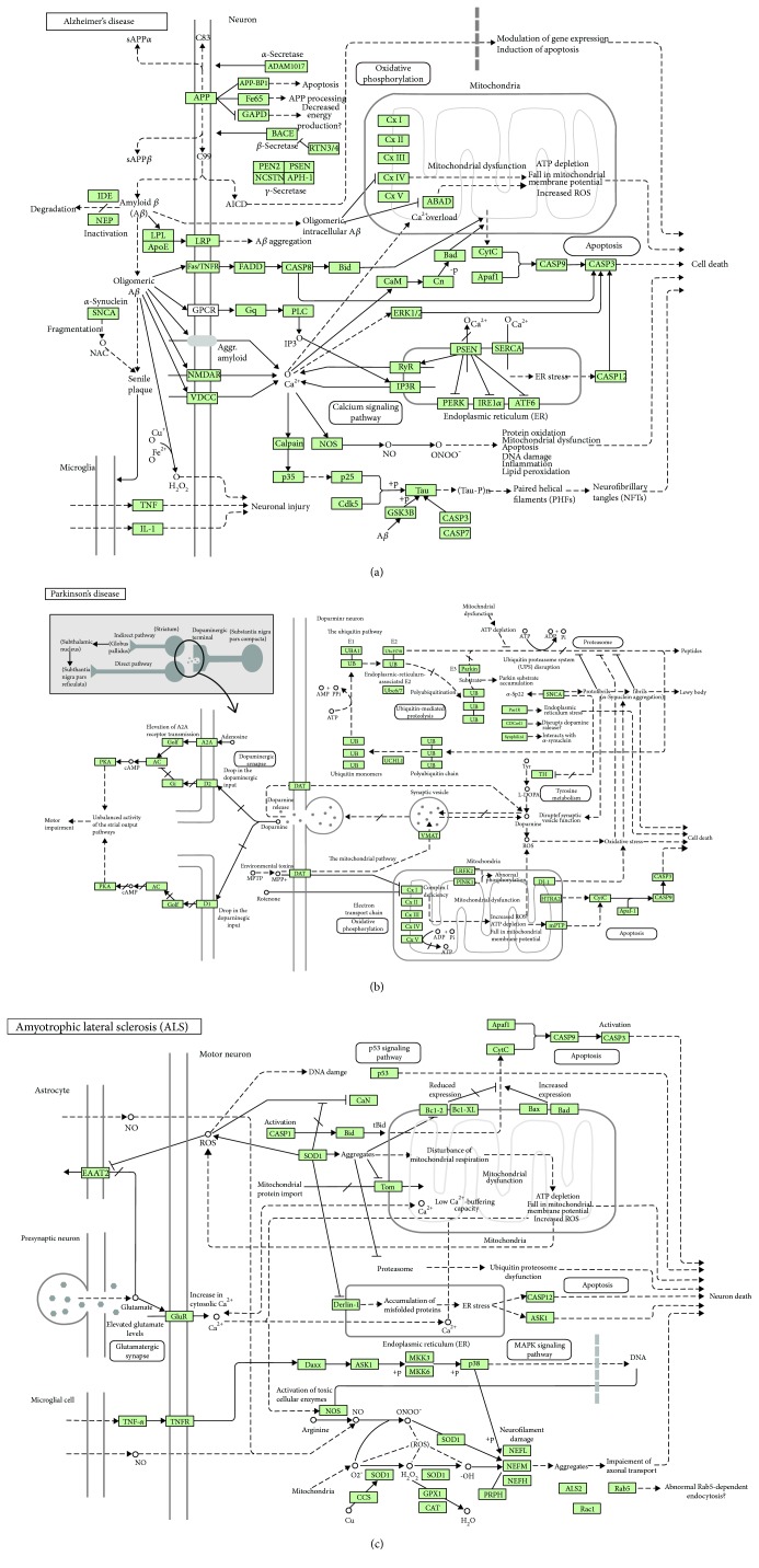
Figure 1.
Schematic KEGG map representations of signaling pathways involved in Alzheimer's disease (a), Parkinson's disease (b), and amyotrophic lateral sclerosis (c). Oxidative stress-induced alterations in signaling pathways, which cause mitochondrial dysfunction, endoplasmic reticulum (ER) stress, and dysregulation of the ubiquitin-proteasome system (UPS) and the autophagy/lysosomal protein quality control machinery, followed by neuronal death, are also shown. Here, → indicates stimulating effects and — indicates inhibitory effects. (a) AD is characterized by the formation of amyloid precursor protein-derived amyloid β-peptide (Aβ), a major component of senile plaques, which forms oligomers to induce pathways initiated by the following receptors: (i) LRP, an apoE receptor; (ii) amyloid precursor protein (APP), an integral membrane protein, mutations of which cause susceptibility to familial AD; (iii) TNF-α receptor (Fas/TNFR) to activate caspases; (iv) GNAQ (Gq)/G-protein-coupled receptor (GPCR) to stimulate phospholipid C (PLC) followed by the activation of inositol-3-phosphate receptor (IP3R) and ER stress; (v) N-methyl-D-aspartate receptor (NMDAR) to cause hyperphosphorylation of tau receptors, and (vi) voltage-gated (dependent) calcium channels (VDCC) followed by neuronal damage through mitochondrial dysfunction and disruption of calcium release from ER. Presenilin 1 and 2 (PSEN1 and PSEN2) proteins belong to γ-secretases that generate Aβ. (b) PD results from the death of dopaminergic neurons in the substantia nigra pars compacta (SNs). Normally, dopamine active transporter (DAT) pumps dopamine out of the synaptic clefts into the cytoplasm. The early onset of PD is associated with mutations in synuclein-alpha (SNCA), ubiquitin carboxy-terminal hydrolase L1 (UCHL1), PTEN-induced kinase 1 (PINK1), leucine-rich repeat kinase 2 (LRRK2), mitochondrial serine protease 2 (HTRA2), parkin, and parkin-associated protein DJ1 involved in oxidative stress. (c) ALS is a lethal disorder characterized by the death of motor neurons in the brain and spinal cord. Mutations in SOD1 may interfere with the neurofilament heavy polypeptide (NEFH) and the translocation machinery, the translocase of the inner/outer membrane (TIM/TOM) that is involved in familial ALS. Proapoptotic THFα acts through its receptor, TNFR, to induce inflammation and apoptotic cell death. The main glutamate transporter protein, excitatory amino acid transporter (EAAT2), is inhibited by ROS produced by mitochondria. Glutamate acts through its receptor (GluR) to increase calcium release from ER and to enhance oxidative stress and mitochondrial damage. Permission 190019 for usage of the following KEGG pathway images was kindly granted by Kanehisa Laboratories [141]: map05010—Alzheimer's disease; map05012—Parkinson's disease; map05014—amyotrophic lateral sclerosis (ALS).

